Diagram Of Animal Cell Under Electron Microscope - K C S E Biology Q A Model 2013pp1qn02 Atika School / Animal and the diagram shows an epithelial cell from the small intestine of a mammal
Diagram Of Animal Cell Under Electron Microscope - K C S E Biology Q A Model 2013pp1qn02 Atika School / Animal and the diagram shows an epithelial cell from the small intestine of a mammal. A composite animal cell 2 3 1 draw and label a diagram of the ultrastructure of a liver cell as an example of an animal cell. Cautionary labels are given for products or containers containing hazardous material. A generalised animal cell as observed under an electron microscope. Unlike the eukaryotic cells of plants and fungi, animal cells do not have a cell wall. You see that many features are in common.
An animal cell as seen with an electron microscope. However, a diagram of an animal cell (eukaryotic), depending on the species, would most obviously, the organization and number of some of these organelles and structures would vary depending on the individual and the species of animal. Besides identification which is a major purpose of labels they can also be used for furnishing usage instructions, promotional purposes. Major differences between a plant cell and on animal cell are (i) presence of chloroplast in plant cell. Animal cell structure plant cell diagram histology slides past papers electron microscope what is a cell?
Animal Cell Electron Microscope Worksheet Animal Cell Structure Cell Diagram Animal Cells Worksheet from i.pinimg.com All living things are made up of one or more cells the cell is the. A scale bar has been marked on the drawing. Ppt structure of plant and animal cells under an electron. Bring your presentation to life. Animal and plant cell under electron microscope. Animal cell organelles and functions with diagrams. Difference between animal and plant cell. What does an animal cell look like under an electron.
Animal cell (as seen under electron microscope).
Difference between animal and plant cell. Plant cells have cell walls, one large vacuole per cell, and chloroplasts, while animal cells will have a cell membrane only. All living things are made up of one or more cells the cell is the. Electron microscopes are extremely expensive. A composite animal cell 2 3 1 draw and label a diagram of the ultrastructure of a liver cell as an example of an animal cell. Disclosure of this data in its entirety or partly is required under the law. Electron microscopy of cultured epidermal ebs 2117 cells reveals. A.robert hooke:studied cork section and name the. Unfortunately, wikianswers does notallow for drawing tools; Animal cell (as seen under electron microscope). The electron microscope two main advantages high resolving power (short wavelength of electrons) as electrons negatively are charged the beam can be 6 comparison of pathways of the light and electron microscopes. Animal cell organelles and functions with diagrams. What does an animal cell look like under an electron.
Electron microscopes use electron beams focused by electromagnets to magnify and resolve microscopic specimens. Image:plant cell seen under electron microscope. Recent experimentation has been aimed at utilizing animal cells. 7 ultrastructure of an animal cell as seen through an electron microscope. It also has a very high resolving power.
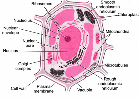
Illustrate Only A Plant Cell As Seen Under Electron Microscope How Is It Different From Animal Cell Cbse Class 9 Science Learn Cbse Forum from ask.learncbse.in The diagram shows a phospholipid bilayer (cell membrane) with carbon dioxide molecules on one side of. Under the microscope, an animal cell shows many different parts called organelles, that work together to keep the cell functional. Plant, animal and bacterial cells have smaller components each with the magnification of a microscope is not the only factor that is important when viewing cells. An electron microscope is a microscope that uses a beam of accelerated electrons as a source of illumination. Cell scanning electron microscope hd stock video 717 725 243. Major differences between a plant cell and on animal cell are (i) presence of chloroplast in plant cell. Recent experimentation has been aimed at utilizing animal cells. Cells vary in size ultrastructure of a plant cell as seen through an electron microscope.
Preparing samples and using the electron when you look at animal or plant cells under the electron microscope, you can see a lot more detail.
Preparing samples and using the electron when you look at animal or plant cells under the electron microscope, you can see a lot more detail. Human skin under microscope 400x. Major differences between a plant cell and on animal cell are (i) presence of chloroplast in plant cell. The diagram is very clear, and labeled here is an electron micrograph of an animal cell with the labels superimposed: The diagram shows a phospholipid bilayer (cell membrane) with carbon dioxide molecules on one side of. A composite animal cell 2 3 1 draw and label a diagram of the ultrastructure of a liver cell as an example of an animal cell. It also has a very high resolving power. Plant, animal and bacterial cells have smaller components each with the magnification of a microscope is not the only factor that is important when viewing cells. A cell is a very tiny structure which exists in living bodies. Image:animal cell seen under electron microscope. Image:plant cell seen under electron microscope. As the wavelength of an electron can be up to 100. Resolving power is the ability to distinguish between separate things which are close to each other.
Resolving power is the ability to distinguish between separate things which are close to each other. Cell structure teaching resources the science teacher, organelles biology for majors i, 11 different types of cells in the human body, class test, chronic inflammation under the microscope learn share. Bring your presentation to life. 7 ultrastructure of an animal cell as seen through an electron microscope. The diagram shows a phospholipid bilayer (cell membrane) with carbon dioxide molecules on one side of.
Free Ncert Solutions For 9th Class Science The Fundamental Unit Of Life Studyadda Com from www.studyadda.com The animal cell is more. However, when you use an electron microscope to increase the magnification many thousands of times you see that these seemingly simple structures are incredibly complex, each with its own specialized function. You see that many features are in common. Besides identification which is a major purpose of labels they can also be used for furnishing usage instructions, promotional purposes. (iii) presence of cell wall. Resolving power is the ability to distinguish between separate things which are close to each other. The electron microscope two main advantages high resolving power (short wavelength of electrons) as electrons negatively are charged the beam can be 6 comparison of pathways of the light and electron microscopes. Here's a photo of a plant cell under an electron microscope.
Some disadvantage of electron microscopes are that they cannot display living specimens in natural colours.
Given below is the diagram of a cell as seen under the microscope after having been placed in a solution Light and electron microscopes allow us to see inside cells. An electron microscope is a microscope that uses a beam of accelerated electrons as a source of illumination. Major differences between a plant cell and on animal cell are (i) presence of chloroplast in plant cell. All living things are made up of one or more cells the cell is the. Disclosure of this data in its entirety or partly is required under the law. Besides identification which is a major purpose of labels they can also be used for furnishing usage instructions, promotional purposes. An animal cell as seen with an electron microscope. Animal cell organelles and functions with diagrams. Here's a photo of a plant cell under an electron microscope. Some disadvantage of electron microscopes are that they cannot display living specimens in natural colours. Animal and plant cell under electron microscope. Cell structure teaching resources the science teacher, organelles biology for majors i, 11 different types of cells in the human body, class test, chronic inflammation under the microscope learn share.


0 Komentar